Posted in Psychology & Medicine

Holistic Medicine

At face value, medicine appears to work on a relatively simple model. You gather information through history taking, clinical examination and investigations such as lab tests and imaging. Then, you narrow down the differential diagnosis to the single most likely diagnosis. Lastly, you treat the diagnosis as per the recommended treatment guidelines.

But if you ask anyone who works in healthcare, they will all know that is not the whole truth. There are so many other factors and variables that play in to the management of a patient that the model above does not address.

For example, you may diagnose a skin infection and prescribe antibiotics, but the person may not have enough disposable income to pay for the medications. You may come up with a plan for the patient to come in to clinic in a week’s time for a review, but they may not have transport or someone to look after their children so that they can come in. You may diagnose that there is nothing medically wrong with the patient, but they may still be worried that they have a serious condition that killed their father.

In medicine, you do not treat the disease; you treat the patient. It is easy to get so focussed on the clinical picture that the overall context is lost. This leads to incomplete care, which causes a variety of issues ranging from patient dissatisfaction to recurrent presentations.

Although it may seem difficult and time-consuming to pay attention to these extra details, it almost always pays off in one way or another. Addressing a patient’s troubled social situation may reduce the number of times they present to hospital, saving significant costs. The doctor taking the time to reassure the patient that their symptom is not concerning for a significant illness may let the patient sleep comfortably at night. Talking through the patient or their family’s concerns and questions might make the worst day of their lives slightly more tolerable.

This approach is useful outside of the hospital too. When you face a problem, regardless of the type, instead of trying to come up with a quick fix to patch it up, try to consider the context of the problem. You may discover that there is a deeper, more fundamental cause of the problem that needs fixing.

Posted in Psychology & Medicine

Laughter Epidemic

It is said that laughter is infectious. In 1962, an extreme case of “laughter infection” happened in village in Tanzania. The phenomenon originated in a boarding school for girls. On January 30, three girls spontaneously burst out in laughter and could not stop themselves from laughing. Soon after, the whole class was suffering from fits of uncontrollable laughter. The “infection” then spread throughout the school, claiming 95 of the 159 students over a stretch of two months. This strange symptom of uncontrollable laughter lasted anywhere from a few hours to 16 days. Interestingly, teachers were not affected and only girls between the ages of 12 to 18 were affected. By March 18, the school was forced to close down due to students not being able to focus during class.

The laughter epidemic was not localised to the school. After the school shut down and the girls returned home, fellow villagers were afflicted by the laughing disease, resulting in 217 villagers being “infected” by May (mostly children and teenagers). By June, the laughing epidemic spread to another nearby school, affecting 48 girls. The epidemic then went on to claim two more schools, forcing them to close down. By the time the epidemic died down (6 to 18 months after “patient zero”), it had affected over a 1000 people and shut down 14 schools.

So what was this strange disease? Was it some new viral infection causing neurological symptoms? Was it a toxin in the water supply? The answer was even simpler: mass psychogenic illness, also known as mass hysteria. Mass hysteria is a psychological phenomenon that occurs in groups placed in high-tension situations, such as within an airplane. This setting is perfect for triggering a mass delusion, causing the person to believe they are suffering from a physical disease. The trigger is usually another “patient” and the hysteria spreads like wildfire, usually by people seeing affected victims. Although the above case makes mass hysteria look like a harmless, amusing phenomenon, psychosomatism (when the mind tricks the body into thinking it is sick) can cause symptoms such rashes, fevers, vomiting and even paralysis. In fact, all of these symptoms were also reported during the Tanganyika laughter epidemic.

Posted in Psychology & Medicine

Trepanation

When and what was the first surgery performed by mankind? Many would believe it to be a simple procedure such as suturing a wound. But would you believe that the earliest surgical procedure was brain surgery in 6500BC? Surprisingly, this is true.

Archaeologists have found a large amount of skulls with a large, round hole in them. Some of the oldest skulls with holes were found in France, where 40 skulls from the Neolithic era were excavated. Archaeologists believed these holes to be from a battle leading to a dent in the skull. However, these holes were actually the results of a surgery (signs of bone recovery can be seen around the edges of the hole, suggesting the patients were alive for some time even after the operation). These skulls all belonged to trepanation patients.

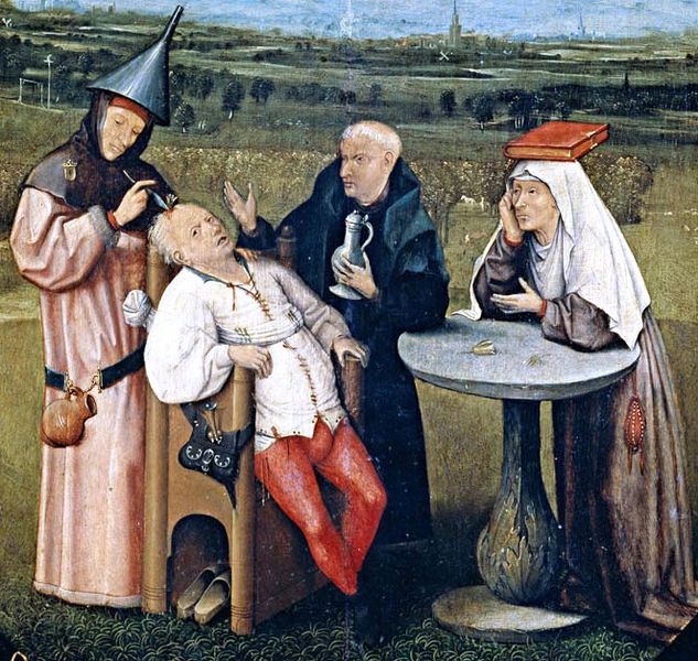
Trepanation is the surgical opening of the skull by drilling a hole in it. This is an ancient surgery that can be found throughout history. Hippocrates and Galen from ancient Greece both recorded detailed instructions on trepanation, ancient Incans performed the surgery and it was also common during the Middle Ages and the Renaissance in Europe. These surgeries were most likely indicated for skull fractures where fragments were embedded in the brain. During the Middle Ages when it was better known that the brain was the seat of the soul, trepanation was used for psychiatric treatments too. For example, in 15th century Netherlands, trepanation was used to excise a so-called stone of madness that was supposedly the cause of insanity. Like this, it was believed that trepanation could release the demons and insanity trapped in the skull.

Although this operation sounds hilariously misled, it is still used in modern medicine. Of course, it is not known to treat insanity, but rather to treat brain bleeds. Extradural and subdural haemorrhages occur when a rupture of an artery in the brain causes a collection of blood in the skull, compressing the brain. This is a dangerous situation which can lead to a stroke or even death. One treatment of this condition is trepanation, or a burr hole, where a small hole is drilled in the skull to relieve the pressure, lowering intracranial pressure and stabilising the patient. Trepanation is an excellent example of how we can learn from the past and how medical knowledge from ancient times is sometimes still valid.

Posted in Psychology & Medicine

Munchausen’s Syndrome

Some people are known to overreact regarding their health, such as a hypochondriac thinking that she has kidney failure because her urine looks slightly frothier. However, some people far surpass the level of hypochondrias to the level of psychiatric disease.
Münchausen’s syndrome patients are known to exaggerate or create symptoms so that the doctor would pay attention to them. When the doctor investigates, treats and sympathises with the patient they gain satisfaction from all the attention they are receiving.
Although this may sound like hypochondrias, Münchausen’s is far more serious.

A Münchausen’s patients are known to cause symptoms just to get attention from others. For example, a common manoeuvre used is the injection of insulin to induce a hypoglycaemic seizure. When their symptoms are “treated”, the patient will most likely invent another factitious disease to be treated for a longer time. They will also seek out many different doctors when the attending doctor catches on to their act. In fact, a Münchausen’s patient will do almost anything to prolong medical care, even accepting unnecessary and risky procedures such as surgeries.

The key difference between Münchausen’s syndrome and hypochondriasis is that the patient is aware that they are not actually sick (hypochondriacs actually believe they are sick). The fundamental basis for Münchausen’s syndrome is the desire for attention. Thus, the main risk factor for developing Münchausen’s is childhood experience of seeing someone close (typically a family member) suffering a debilitating disease. For example, if a girl sees her sister suffering from leukaemia and receiving all the attention of everyone around her, she may develop feelings of jealousy and later try to duplicate the scenario. As a patient, the person feels safe and comfortable and this feeds their addiction to medical care.

As Münchausen’s patients are very proficient liars and act completely like an actual patient, doctors must rule out any diseases before suspecting that their patients have a psychiatric problem. However, some signs such as the patient being overly keen on receiving procedures such as biopsies or continuously developing random symptoms may indicate Münchausen’s.

Interestingly, a similar condition called Münchausen’s syndrome by proxy also exists, where a caregiver (e.g. mother) convinces a doctor that the person they are caring for (e.g. child) are sick. Unfortunately, as these patients actually cause illness in the child, it is considered a form of child abuse. Common “symptoms” include: growth problems, asthma, allergies, vomiting, diarrhoea, seizures and infections. This may lead to the child developing Münchausen’s syndrome in the future.

Posted in Psychology & Medicine

Korsakoff’s Syndrome

It is a well-known fact that excessive drinking leads to a so-called “blackout”. This form of memory loss is common in normal people and cannot be seen as a major illness. However, there is another disease that can be caused by excessive drinking called Korsakoff’s syndrome. Strictly speaking, this is not caused by alcohol but due to a thiamine (vitamin B1) deficiency and is commonly found in alcoholics and malnourished patients (it has also been reported to be caused by mercury poisoning and after centipede bites in Japan).

The six characteristic symptoms of this syndrome are: anterograde (cannot form new memories) and retrograde (cannot remember old memories) amnesia, confabulation, lack of detail in conversation, lack of insight and apathy.

Korsakoff’s syndrome patients show a very peculiar behaviour. As stated before they suffer from both anterograde and retrograde amnesia so not only can they not remember the past but they cannot make new memories either. Ergo, the brain uses information from its surroundings and attempts to recreate the lost memories, the result being confabulation. Confabulation is essentially what happens when the brain tries to fill in blanks in memories with false information. Confabulation is seen in everyday life too with healthy people but in the case of Korsakoff’s patients the effects are significantly more profound. For example, if you ask a patient what she did yesterday, she may look at your horse-print tie and claim she was horse-riding. If you ask the same question an hour later without your tie and instead holding a book with a photo of a Ferris wheel on the cover, she’ll state that she was at the amusement park. As one of the leading causes of amnesia and confabulation, Korsakoff’s should be suspected in any alcoholic or very underweight patient who keeps changing their stories around. 

As previously explained, the disease is caused by thiamine deficiency – therefore, the treatment is administering thiamine. But if the syndrome has persisted for a long time, the brain injury may be permanent. Also, treating the underlying alcoholism and malnutrition is important. 

If the thiamine deficiency is prolonged, it may lead to another disease called Wernicke’s encephalopathy. This is known as Wernicke-Korsakoff’s syndrome and in addition to the above symptoms, the patient may also experience confusion, tremors, nystagmus, paralysis of eye muscles, ataxia, coma and can eventually lead to death. All because of a deficiency of a single vitamin.

Who said nutrition is not important?

image

(NB: Dory from Finding Nemo is one of the most accurate portrayals of amnesia in films)

Posted in Psychology & Medicine

Couvade Syndrome

A patient comes to a doctor complaining of the following symptoms: “I can’t sleep because my teeth have been aching for the past few weeks. My head is killing me and whenever I wake up in the morning my stomach hurts and I feel nauseous and want to vomit. Sometimes I have no appetite and sometimes I crave a certain food. My breasts have gotten bigger and my stomach is bulging quite a bit.”
The patient has been married for three years and in a few months will have a beautiful daughter.
What is the diagnosis?

Most people would immediately say “Pregnancy!”. But there is one small detail that was left out: the patient is a man.
It is common sense that men cannot be pregnant. So what is this man suffering from?

Couvade syndrome is also known as sympathetic pregnancy. In other words, the husband subconsciously copies his wife’s pregnancy and suffer the same symptoms. This syndrome can be severe enough to cause labour pains, nosebleeds and even post-partum depression.
The cause has not been established, but it is likely to be psychosomatic, where psychological symptoms are expressed through physical symptoms, or due to changes in hormone levels.

Posted in Psychology & Medicine

Gait

In medicine, a person’s way of walking is termed gait. By analysing a person’s gait, a trained professional can gain insight into what pathologies the person may be suffering from. For example, just from the way the patient limps, the doctor may discover that the patient has an incurable degenerative brain disease.

The most common gait abnormality is the antalgic gait, or limping due to pain. Most people would have experienced the difficulty of walking with a sprained or broken ankle, muscle ache or knee problems. This is easy to spot as the patient quickly switches to the other feet when leaning on the affected leg due to the pain. Therefore, the side that stays on the ground less than the other is the affected leg.

Sometimes, you can see a person “waddling” along as they swing from one side to the other. This may be a waddling gait, also known as Trendelenburg’s gait, caused by a weakness in the hip muscles that support the pelvic girdle, either due to muscle or nerve damage. As the patient cannot support their weight on the affected side, their pelvis tilts towards the opposite side. To avoid falling over, the patient lurches their body towards the other side, causing them to waddle. Looking at the tilt and lurch gives insight into what side is affected.
Another rather common gait is the steppage gait, where the person lifts one leg higher than the other, while their foot drags on the ground. This is caused by nerve damage leading to the loss of ability to lift the foot up (termed foot drop).

As the brain controls the motor system, damage to the brain also leads to motor dysfunction. A common example is a stroke.
If the stroke damages a significant part of the motor cortex, the patient suffers from hemiparesis/hemiplegia, or weakness/paralysis of one half of the body. This causes the limbs on the affected side to stiffen, as seen by an extended leg pointing inwards and retracted arm. The patient has to swing the affected leg around while they walk as they cannot flex the hip, known as a hemiplegic gait.
If the cerebellum is damaged, balancing becomes an issue. This causes the patient to suffer from ataxia, where they cannot coordinate their movements and are prone to toppling over. These patients tend to sway violently from side to side as they try to walk in a straight line.

Lastly, degenerative brain diseases can also affect gait. There are two main examples.
In Parkinson’s disease, the patient suffers from what is called hypokinesia and bradykinesia – reduced and slow movement. This leads to a shuffling gait where the patient walks slowly by shuffling their feet in small steps. They are also stooped over and are often seen with a pill-rolling tremor of their hands – a cardinal symptom of the disease.
In Huntington’s chorea, the opposite (hyperkinesia) occurs. This causes flailing as the muscles contract in an uncoordinated manner, including both the arms and legs. Ergo, their gait is quite jerky and interrupted by bouts of flailing, termed choreiform gait, but their balance is fine so walking in a straight line is still possible.

Knowledge of these disorders may help one appreciate the suffering a patient walking along the street has to undergo everyday of their life.

(Video demonstrations: http://stanford25.wordpress.com/gait-abnormalities/)

Posted in Psychology & Medicine

Diagnosis

A man comes in to a clinic and tells the doctor that he is in great pain.
It hurts if he prods his arm with a finger. The same applies to his leg and his torso.
He says that after looking on the internet, he is sure that he has fibromyalgia syndrome, and asks to be prescribed some painkillers. Also, he is worried that it could be necrotising fasciitis and demands a biopsy and antibiotics.

The doctor, without saying a word, gently grabs his hand, then squeezes his index finger. The man feels a shooting pain and screams in agony.

The diagnosis? A broken finger.

Posted in Psychology & Medicine

CPR

CPR stands for cardiopulmonary resuscitation – or in plain English, artificially (and partially) restoring the function of the heart and lungs of an unconscious, pulseless person. As blood flow (perfusion) is critical in the survival of major organs such as the brain, this procedure can save lives by prolonging a victim’s life until the paramedics arrive to provide professional medical care.

When the heart stops beating, or becomes inefficient due to erratic beating, blood flow stops. In the case of the brain, this means that the cells will start dying after 4~5 minutes if perfusion is not restored. CPR can restore about 30% of perfusion, delaying the onset of brain death.

This may be critical when someone suffers a heart attack (myocardial infarction) and paramedics will not arrive for over 10 minutes. Ergo, this is one of the most important emergency skills one should know to help people in need as soon as possible.

There are different guidelines for CPR in many countries, but here is a standard procedure guideline (NZ).
It is summarised into the acronym: DR’S ABCD (doctor’s ABCD), and is a flowchart that goes from one step to the next (detailed explanation after summary).

  1. Danger: check that area is safe and risk-free
  2. Response: check for patient response by shouting, shaking, pain
  3. Send for help: pick one person to call emergency services
  4. Airway: check airway, remove obstruction, tilt head back and lift chin
  5. Breathing: check for breathing, go to CPR if no breathing
  6. Circulation: check for pulse if breathing, if no pulse, start CPR (30 chest compressions : 2 breaths)
  7. (Defibrillation): follow AED instructions

The first rule of first aid is that you must not put yourself in danger. For example, if the patient is on the road, pull them to a safe area to minimise the risk to your own health.

Then, check for a response. The easiest way is to call loudly to them such as “Can you hear me”, and inflicting pain (such as rapping on their chest or shaking their shoulders) and see if they become conscious.

If they remain unconscious, immediately designate a person around you by pointing to them (otherwise they will be less likely to be responsible) to call the emergency service (111, 911, 119 etc.), alerting them the location and state of the patient.

This is the point when clinical skills come in.

Airway: An unconscious person may have their airway obstructed by vomit or their own tongue (which falls back by gravity into the throat). You must secure the airway by scooping out any material, and clearing the tongue out of the way. This is done by tilting the head back far (as if they are looking up), then using one hand to pull their chin out. This opens the airway up so that mouth-to-mouth becomes effective.

Breathing: Put one ear right next to the person’s nose and mouth and check for any breathing sounds or air flow. If they are breathing, check the pulse to see if they are pumping blood. If not, go straight to CPR.

Circulation: It is best to check the central pulses such as the carotid (side of neck, next to the Adam’s apple), brachial (squeeze inner side of biceps) or femoral pulses. The carotid is often the easiest as most people know how to take it. If you feel a pulse, put the patient in recovery position as they are just unconscious, breathing and has blood flowing. If not, proceed to CPR (as you do with when the patient is not breathing).

CPR is composed of two actions: chest compressions and mouth-to-mouth breathing. The former is the strong compression of the chest wall to squeeze blood in and out of the heart; the latter is breathing air into the patient’s lungs and letting exhalation come out naturally.

Chest compressions are often misrepresented in medical dramas, and is extremely important that you do it correctly. First find where the sternum is (centre of ribcage, between the nipples) and place the heel of your left palm on it, then spread your fingers out. Put your right hand over your left and close your fingers around it for a good grip. If the patient is lying flat on the ground (with head tilted back), kneel beside them and stoop over their chest with straight, locked arms (bent arms exert much less pressure).

You are now ready to begin chest compressions. Press down hard, until the chest wall is compressed to about 1/3~½ depth (the chest wall is a springy structure, and do not worry about broken ribs, as being alive is more important for the person), then ease pressure to let it bounce back up. Ideally the time pushing and the time letting it bounce back should be the same, giving a good rhythm. Repeat this 30 times at the beat of 100/min, or in easier terms: to the beats of the Bee Gee’s song Stayin’ Alive (scientifically proven).

After 30 compressions, tilt the patient’s head back, lift their chin up, and lock your mouth over their mouth and nose to make an airtight seal. It is crucial that you use a face shield to prevent the spread of disease. Be aware that breaths are less important than the compressions, so if you do not have a face shield, let someone else do the breathing and focus on chest compressions. Pinch the nose closed to ensure air does not escape.
Forcefully breathe into them and look for the chest rising. Let go of the nose and pull away so that they can breathe out. Repeat once, then return to chest compressions.

After 2 minutes of CPR (30 compressions : 2 breaths, repeat 4 times), change places with another person capable of CPR, as otherwise you will tire out and become inefficient.

Defibrillation is only possible if you are near an AED (automated external defibrillator). Nowadays, AEDs are designed to be completely user-friendly so simply follow the instructions on the machine.

It is important to note that not all abnormal heart rhythms are “shockable” (see Flatline). Follow the AED’s instruction, as it will state whether shock is advised or not. Make sure that CPR is still happening continuously.

Repeat until help arrives.

As a final note, remember that the patient is dead whether you do CPR or not, so there is nothing to lose. Believe it or not, this will be of incredible help in calming your mind when struck with such an emergency. Even with CPR, there is a maximum 30% chance the patient will survive, 10% if it occurs outside the hospital. But if you do nothing, their survival chance will be 0%, so put all your energy into resuscitating them, and you may just save a life.